ANKLE PROSTHESIS
Ankle replacement is the solution for the ankle arthritis.
- My Experience
- Ankle Arthritis and Quality of Life
- Post-Traumatic Ankle Arthritis
- Ankle Fractures and Ligament Injuries
- The History of Ankle Arthroplasty: Fix-Bearing and Mobile-Bearing
- Ankle Arthroplasty Today
- Resurfacing Ankle Prosthesis
- Ankle Replacement and Learning Curve: The Value of Numbers
- Our Operating Room
- Ankle Arthroplasty: The Postoperative Route
- The Value of Teaching
- Scientific Research and Ankle Replacement: Our Results
- The future is now: Weight-Bearing CT scans, robotics, augmented reality, and ankle prostheses
- FAQ (Frequently Asked Questions)
My Experience

Duke University
When in 2008, at Duke University, I began to increase my interest of ankle prosthesis, we were still at the beginning of this surgical solution.
In that moment there was talk of isolated experiences; good results in the surgeons’ designs of the implant and mixed results in the rare cases of independent surgeons.
Current solutions were not yet able to be the answer for patients suffering from ankle arthritis since prostheses were still looked at with fear and doubt. The procedure known as ankle arthrodesis was looked at favorably, especially in cases involving young patients and high functional request.
What does arthrodesis mean? It is a surgical procedure that involves the fusion of the ankle in a favorable position to allow the development of a walk by taking advantage of the movement of nearby joints.
It is a solution that we have extensively studied in the past, grasping its weak side, in the alteration of the natural walk and the consequent arthritic degeneration of nearby joints.
Ankle Arthritis and Quality of Life
If we were to analyze the characteristics of a patient suffering from ankle arthritis, we would be shocked by the significant negative impact this condition has on their quality of life.
The word “arthritis” is often associated with older age. We picture an elderly patient, reliant on a cane or crutches to manage even short distances. We think of someone for whom simple daily activities, like putting on trousers, tying shoes, or getting in and out of a car, become difficult.
These patients often find themselves having to rely on painkillers and anti-inflammatories just to get through their day. This approach can lead to serious long-term issues with other organs involved in drug metabolism, such as the liver and stomach.
The situation is even more challenging for those with ankle arthritis, as they are typically much younger patients, often at the peak of their social and professional lives. For these individuals, daily life becomes a constant struggle, and work can become impossible. Depression is a serious risk.
Post-Traumatic Ankle Arthritis

Most of my patients are under 50 years old and have a history of fracture trauma that occurred 5 or 10 years before I met them.
Sometimes the interval between the injury and the development of arthritis is shorter. They are patients, as mentioned, at the height of their lives, who have had accidents:
- riding motorcycles;
- skiing;
- working on a construction site;
- repeated distortion traumas found with ex-soccer players, volleyball players, or basketball players.
Alternatively, it also involves even more unfortunate patients suffering form systematic inflammatory diseases, such as rheumatoid arthritis, systematic lupus eritematosus, or intra-articular hemoglobin deposition diseases, such as hemophilia and hemochromatosis.
They are subjects who often have poor quality bone tissue and serious functional limitations due to the chronic therapies they must undergo.
Mark Glazebrook (a Canadian colleague) in the “orthopedic bible”, the scientific journal “JBJS Am” (2017), recently quantified that the disability of patients suffering from ankle arthritis is superior to the disability caused by knee arthritis and is equal to that of hip arthritis.
This is not a competition for the most misfortunes and diseases, but a way to quantify the serious problems faced by patients with ankle arthritis and to clinically motivate the importance of early and priority treatment.
Ankle Fractures and Ligament Injuries
The ankle is an extremely congruent joint, with its surfaces fitting together as perfectly as the pieces of a puzzle. It is such a finely balanced system that, when undisturbed, it remains durable over time.
The ankle is a joint with an intrinsic stability. The role of the ligaments takes over for dynamics or during movement.
Repeated ankle injuries can lead to ligament and cartilage damage. The onset of arthritis can be prevented with early treatment of instability, which involves reconstructing the cartilage and repairing the injured ligaments.
Fractures can affect the tibia, fibula, malleoli, and talus.
In these cases, "acute early treatment" aims to restore the original anatomy. It is the first step in preventing osteoarthritis, which can still develop even after a successful anatomical reconstruction.
The role of the fibula is often underestimated in fracture treatment. It’s not uncommon for patients to undergo perfect surgery for the tibial fracture, but the original length of the fibula may not be restored. In addition to pain and disability, these patients may develop a valgus deformity if the fibula is too short, or a varus deformity if it is too long.
Another aspect to consider is translational deformities. These occur when the talus (the bone in the foot that forms the ankle) becomes unstable and "slides" forward relative to the tibia and its center of rotation. This leads to a significant reduction in ankle movement. Patients with this deformity often limp noticeably, keeping the affected limb stiffly on tiptoe, with the knee in a position of "recurvatum."
In these cases, it is important to correct the translational deformity rather than opt for an "anterior release" with arthroscopic treatment, as was once common.
In fact, patients I see for the first time often show significant worsening if they have previously undergone ankle arthroscopy elsewhere.
Arthroscopy is a procedure in which the joint is inspected and cleared through two small incisions.
It is particularly useful in cases of cartilage lesions because it avoids "opening" the joint, thereby reducing the risk of post-surgical stiffness.
However, it proves to be ineffective and sometimes counterproductive in cases of impingement or reduced movement caused by the formation of anterior exostosis.
In these cases, the surgeon’s goal should be to restore the correct axis of rotation of the joint.
This is a cutting-edge concept and has become a “hot” topic, which my team brought to the forefront of scientific discussion with our study, recently published in the European Society of Ankle and Foot Surgery (EFAS) journal.
The History of Ankle Arthroplasty: Fix-Bearing and Mobile-Bearing

Ankle prosthesis with mobile bearing has become a reliable solution for treating ankle arthritis.
The excellent results we see today are part of recent history. In fact, there has been an unstoppable evolution in design and tribology (the study of materials and their interaction) over the past twenty-five years. This progress is the result of intense scientific activity, with two major schools of thought: American and European.
The Americans developed extensive prosthetic designs that are anchored to the talus and tibial surfaces. These prostheses consist of two components, with movement between them (Fixed-Bearing).
While these designs offer the advantage of providing a good range of motion for the prosthetic ankle, they come with the disadvantage of being subjected to significant mechanical stress at the bone interface. I encountered these prostheses during my time working in the United States and, frankly, I observed more long-term disadvantages than advantages with them.
The winning prosthetic concept comes from Europe and is known as the Mobile-Bearing prosthesis.
To reduce the immense stress placed on Fixed-Bearing prostheses, a Danish surgeon, H. Kofoed, was the first to propose a revolutionary three-component design: a tibial component, a talus component, and a mobile polyethylene pad, referred to as a meniscus, to dissipate the stress between the two main components.
This concept gained widespread popularity and was modified by several authors. The most successful version is likely the "Hintegra" prosthesis, which I was one of the main users of in Italy. It was designed by Beat Hintermann, one of my mentors during my training and someone with whom I worked in Switzerland.
The advantages of this prosthesis included its smaller dimensions, which preserved the patient’s bone, and osseointegration that did not require cementation.
The main disadvantage, however, was the relatively high rigidity of the system compared to Fixed-Bearing models.
Ankle arthroplasty today
Today’s ankle prosthesis is the gold standard treatment for symptomatic ankle arthritis.
It enables the patient to maintain a natural walking pattern without the need for adaptations that could overload nearby joints. It is also compatible with low-impact sports for younger, more active patients.
It is now clear what the key ingredients for a successful replacement are: preserving bone stock and ensuring proper alignment at the end of the procedure to guarantee stability and function. All successful prosthetic designs prioritize bone preservation.
This focus on preserving bone paved the way for the development of "resurfacing" procedures.
Resurfacing Ankle Prosthesis
.webp?width=1200&height=675&name=protesi-di-caviglia-fix-bearing-desktop-min.jpg%20(1).webp)
Resurfacing refers to a prosthesis that closely mimics the natural ankle and, when implanted, minimizes bone sacrifice.
In essence, resurfacing is a system that aims to recreate the original shape of the ankle, from the preparatory cuts to the insertion of the prosthesis.
This is a crucial feature because it provides the patient with a second chance. The more bone the surgeon can preserve, the greater the opportunity for future repairs once the prosthesis has worn out.
Ankle Arthroplasty and Revisionary Repairs
It's important to remember that, in the case of the ankle, arthritic patients tend to be younger and more active than those suffering from arthritis in the hip and knee. The possibility of performing a revisionary repair is undeniably a safety factor.
Design plays a crucial role in the success of an implant, but it must be complemented by material choices that enhance these advantages.
Modern prostheses focus significantly on the osseointegration process. Historically, prostheses were coated with hydroxyapatite to promote bone metabolism. Today, the most reliable approach for integration is the use of trabecular metal, a material derived from Tantalum processing, which closely resembles bone.
In practice, bone cells (osteoblasts) "recognize" and "inhabit" the trabecular metal, integrating it as if it were bone itself. This process eliminates the need for cementation procedures.
Ankle Replacement and Learning Curve: The Importance of Experience
Ankle prosthesis is the preferred treatment for ankle arthritis.
Like many procedures in orthopedics, it is burdened by a significant "learning curve" for the surgeon. Just like any other professional, a doctor needs to learn and refine their skills with each procedure. This is especially important for a joint like the ankle, where the number of surgeries performed is relatively low.
This aspect has always been a priority for my team, which has been a strong advocate for ankle prosthetic surgery. We even published an insightful article on the subject in the European Ankle and Foot Surgery Journal.
We studied the surgeon’s learning curve in approximately thirty cases of ankle prosthetic surgery. This research underscores the importance of reference centers in providing high-quality care for patients suffering from a relatively rare condition like ankle arthritis.
These numbers should not be seen as strict boundaries, but rather as encouragement for those of us who, like our team, are committed not only to patient care but also to training fellow professionals across Europe and around the world.
Today, with over one hundred cases of the "Hintegra" prosthesis (Mobile-Bearing) and more than three hundred cases of the TM-Ankle Zimmer-Biomet prosthesis (Fix-Bearing or lateral access resurfacing), we have developed a unique approach to the treatment of ankle osteoarthritis.
The evolution of our methods and results has led us to define and refine our original surgical technique, which we published in the international scientific journal Sicot-Journal.
This technique is now recognized as a benchmark for ankle prosthesis with a lateral approach.
Our Operating Room

These numbers have allowed for the careful evolution of our technique.
I am privileged to have trained a group of professionals who are now leaders in their field.
I am privileged to have trained a group of professionals who are now leaders in their field.
Dr. Indino, Dr. Maccario, and Dr. Manzi are my team. They have grown alongside me, and today, they are far more than just skilled surgeons. When I see them working, I know they will always be in the right place at the right time. This brings me great peace of mind, even in the most challenging situations.
The operating room is not just about scalpels and surgeons; it’s about organization and technology. The real leap in quality comes from making everything reproducible, fluid, and coordinated.
Surgeons who visit us from all over Europe are often amazed by the level of organization here. We have two operating rooms dedicated to minimizing downtime, a recovery room for immediate post-surgery care, and a team of dedicated nurses, all part of the Casco Team. We couldn’t treat so many patients with such consistent results if we were working alone.
When a patient thanks me fifty days after surgery, walking on their own, it’s a testament to the entire team’s efforts!
Ankle Arthroplasty: The Post-Operative Journey
After a procedure that typically lasts under ninety minutes, the patient is transferred from the operating room to the recovery room, where two nurses and an anesthesiologist oversee immediate post-operative care.
The patient is fitted with a fiberglass boot (extending below the knee), which is applied in the operating room and will remain in place for two weeks. The operated limb will stay anesthetized for an extended period, often throughout the night. Blood loss is minimized, so routine blood transfusions are not necessary.
The following day, the ankle will be medicated through a lateral opening in the plaster, which was made during the surgery.
Post-Operative Recovery After Ankle Arthroplasty
Within two days, most patients who have undergone ankle surgery experience good pain control and are ready for discharge.
The first follow-up appointment is scheduled for fifteen days post-surgery, during which the sutures and plaster will be removed, and the patient will be given a walking boot, which can be removed at night. This boot will remain in place for the next three weeks as part of the recovery process.
At five weeks after surgery, the patient can begin to bear weight on the foot without the boot. Crutches can be discarded, and the patient can gradually resume normal activities, including driving.
At this point, physiotherapy focused on weight-bearing recovery will begin, often supported by hydrokinesitherapy (walking in water), along with strengthening and stretching exercises for the triceps.
The value of teaching
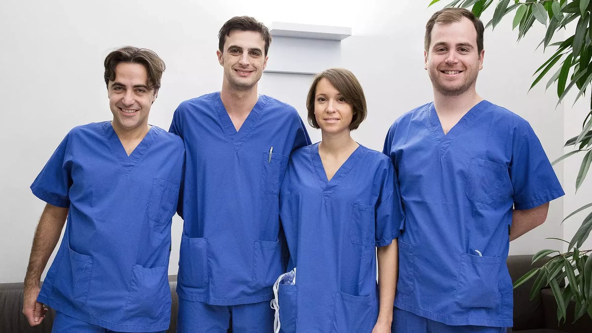
Almost every day, we welcome visitors, not just from Italy, but from all over Europe. We have hosted surgeons from countries such as France, England, Ireland, Belgium, the Netherlands, Germany, Spain, Poland, Russia, and China. These colleagues are interested in learning about our approach to treating ankle osteoarthritis.
Because we firmly believe in the quality of our work, I felt it was important to guide these surgeons on their journey into arthroplasty, offering support and advice to help them in their practice.
This often takes me to operating rooms across Europe, where I guide these colleagues as their mentor during their first surgeries. I contributed to the first ankle prosthesis with lateral access in Spain, the first in Belgium, and the first revision with a lateral approach in Poland.
In terms of numbers and scientific activity in Europe, we are changing the paradigm that young Italian surgeons need to go abroad to learn ankle surgery. To support this, we have developed fellowship and super-specialization courses, open to Italian, European, and Asian colleagues, which have generated significant interest.
A fellowship is a post-specialty training program in which we provide both professional training and financial support. It offers young, enthusiastic surgeons the opportunity to gain exposure to this surgery, which is still evolving in many parts of the world. For me, it’s also a chance to be surrounded by enthusiasm, curiosity, and an added boost to our research efforts.
Scientific Research and Ankle Replacement: Our Results
When we adopted the concept of the lateral approach, fibula osteotomy, and the new prosthetic resurfacing, it truly felt like a revolution.
The primary motivation was to gain direct access to the center of rotation of the ankle, which had remained hidden and inaccessible with the anterior approach.
Today, it is the most widely used technique in Italy for many reasons. Most of the advancements in this area are directly linked to our scientific team.
Dr. D’Ambrosi and Dr. De Silvestri dedicate themselves wholeheartedly to the continuous gathering of data in prosthetics and cartilage reconstruction.
Their work, along with our international collaborations, has enabled us to achieve remarkable results in a very short time, exactly as scientific progress in this field should be: efficient and responsive.
We initially shared the findings of our studies at national and international conferences, including SIOT (Italian Society), AOFAS (American Society), and EFAS (European Society).
We later published our findings in the leading scientific journals of our field: Foot and Ankle International (the scientific journal of AOFAS) and Foot and Ankle Surgery (the scientific journal of EFAS).
We believe the lateral approach enables the surgeon to address one of the most significant negative changes that occur in arthritic ankles: the retraction of the posterior soft tissue.
In our hands, this approach provides a more effective and stable correction of post-traumatic deformities over time compared to other techniques.
One of our most significant studies involved comparing healthy ankles, ankles with a mobile-bearing prosthesis (third generation with an anterior approach), and ankles with prostheses using a lateral approach.
Through rigorous radiographic analysis, we demonstrated that the curved cuts and lateral approach are more effective in restoring an ankle that closely resembles a healthy one, with remarkable results in terms of movement, a natural gait, and “proprioception” (the patient's sense of having their ankle back).
Another pioneering study examined the efficiency and reliability of ankle joint surgery combined with additional procedures for deformity reconstruction, such as subtalar arthrodesis.
This experience has paved the way for using prosthetics in more complex cases of arthritis that require surgery on multiple skeletal segments, ultimately achieving the goal of a functional, aligned, and stable foot.
The future is now: Weight-Bearing CT scans, robotics, augmented reality, and ankle prostheses
Surgery is not a heroic or romantic act, but rather a privilege for the surgeon—an opportunity to help others and connect with them. It is not a matter of improvisation; instead, it is the result of a well-organized team effort and meticulous planning. These tools, like surgery itself, must be understood, applied, and continuously developed. They require a learning curve for the team using them. Most importantly, they demand dedicated resources for this new technology, including the formation of a specialized team focused on foot and ankle treatment.
Today, we have tools that, when combined with augmented reality and robotic surgery, provide—and will continue to provide—greater reliability for surgeons in the operating room. One such tool is Disior (Bone-Logic application), which enables us to plan and visualize in advance the impact of corrections on the foot-ankle complex. In other words, it allows us to preview the procedure before it is performed in the operating room.
This revolutionary approach is made possible by the innovative cone-beam CT scan technology. It allows for a CT examination to be performed while standing, with a radiation dose comparable to that of an X-ray and significantly lower than that of a traditional CT scan. This marks a paradigm shift, bringing three-dimensionality to the preoperative analysis of the foot and ankle.
The impact of these innovations is particularly evident in the treatment of ankle arthritis, which is most often post-traumatic and frequently accompanied by deformities. These deformities must be corrected during ankle replacement surgery to ensure the prosthesis functions properly. If the goal is proper alignment and mobility, modern, precise planning is essential.
Weight-bearing X-rays were the first tool used to assess deformities and plan realignment. To complement this, we always relied on CT scans to evaluate bone quality and three-dimensional structure. However, traditional CT scans lacked the ability to capture images under load—a limitation that has now been overcome with new technology.
FAQ (Frequently Asked Questions)
Ankle arthritis: is a prosthesis the only solution?
I’ll answer with a quote from one of my mentors, Prof. Beat Hintermann:
"I am happy for the successful ankle replacements I performed to avoid fusion, but I am proud of the successful joint-preserving surgeries I did to avoid ankle replacement."
This means that the primary goal of a team dedicated to treating ankle arthritis should be to preserve the joint, correct deformities, and regenerate cartilage whenever possible. When joint preservation is no longer an option, the focus must shift to maintaining function by implanting an ankle prosthesis.
What is the difference between an ankle prosthesis and arthrodesis?
Arthrodesis is a surgical procedure that fuses a joint. In the case of the ankle, it is considered when the joint cannot be preserved or when a prosthesis is not a viable option. However, it comes at the cost of sacrificing movement.
In the past, arthrodesis was regarded as the definitive solution for treating ankle arthritis. Today, in specialized centers where dedicated surgical teams perform a high volume of procedures (at least 30 per year), prosthetic surgery has become the gold standard.
An ankle prosthesis preserves joint mobility. To minimize the risk of complications—such as infection (<2%) and loss of correction (<2%)—it is crucial that the procedure be performed in a specialized center.
As a result, the modern approach to treating ankle arthritis focuses on centralizing care in high-volume reference centers. These centers maintain a Prosthesis Registry, utilize the latest technology, and provide patients with the highest standard of treatment.
Does ankle arthrodesis wear out? What about a prosthesis?
Ankle arthrodesis sacrifices the joint, which increases stress on the surrounding joints, particularly the knee and foot. While a patient with ankle arthrodesis is not at risk of wear on the ankle itself (since it no longer exists!), they are more likely to experience increased wear on adjacent joints, potentially requiring salvage surgery.
A prosthesis, on the other hand, does carry a risk of revision. However, advancements in materials and a deeper understanding of the condition have significantly improved implant longevity. In specialized centers, where experienced surgeons have completed their learning curve, prosthesis survival rates in the medium to long term are now comparable to those of knee replacements.
What is a prosthesis made of?
Most prostheses are made of a combination of metal and polyethylene.
The metal components can be cobalt-chrome (with or without a hydroxyapatite coating to promote osseointegration), titanium, or porous titanium.
The prosthesis I use most frequently—the Resurfacing TM Ankle—is the only ankle prosthesis featuring a metal-back (the part that interfaces with the bone) made of Trabecular Metal, an alloy derived from processed tantalum. It also incorporates cross-linked polyethylene, a high-density, wear-resistant material.
What does the Fast-Track Rehabilitation protocol for ankle prosthesis involve?
This protocol includes immediate weight-bearing and gait re-education (walking) starting from the first post-operative day.
During hospitalization, the patient undergoes Game Ready therapy—the most advanced technology for continuous cold exposure—and magnetotherapy (Lympha-therapy). Additionally, they receive training on how to walk properly.
The patient is discharged without a plaster cast and with instructions to bear full weight and begin walking immediately, as taught during their hospital stay.
This protocol, developed by my team, is designed to optimize the recovery of patients with high functional demands.
Do plates and screws need to be removed before prosthetic surgery, or can they be removed during the same procedure?
Technically, it is possible to remove the fixation devices (plates and screws) during the same procedure as prosthetic surgery.
However, data from our Prosthetic Registry and scientific studies published in leading journals have provided a clear guideline: it is preferable to remove the fixation devices in a separate, preliminary procedure if a plate is present.
This significantly reduces the risk of infection and leads to better outcomes in prosthetic surgery. Removing them earlier increases the overall safety of the procedure.
