What is the Achilles Tendon?
Its task is to transmit the force generated by the triceps surae to the entire foot and ankle complex.
Therefore, it is the tendon that must withstand the greatest mechanical stresses of the entire body.
In order to perform this function efficiently, it has an important structure for controlling both its own function and the distribution of loads (proprioception).
What are the main functions of the Achilles Tendon?
The Achilles tendon is thus a means of transmitting and controlling forces that contributes, when intact, to:
- stability
- movement
- balance
Why is it so important to take care of it?
Taking care of the Achilles tendon means paying attention to:
- the muscle tone of the triceps surae, by training it
- the myotendinous tension, through stretching and proprioceptive work
In particular, learning to listen to all the neuro-muscular spindles—those responsible for producing force as well as those responsible for control—means increasing our control abilities, learning to manage muscle fatigue, and understanding when we are approaching our limits.
It means preventing Achilles tendon injuries, but also preventing falls in general and preserving our motor abilities overall.
Achilles Tendon Conditions: Achilles Tendinopathy
The Achilles tendon can be affected by several conditions.
Some are directly related to Achilles tendinopathy, while others involve the other muscles that form part of the Achilles complex.
Here are the main Achilles tendon disorders:
- Insertional Achilles tendinopathy
- Non-insertional Achilles tendinopathy
- Muscle injuries: calf strain or tear
- Myotendinous injuries: Achilles tendon tear
- Achilles tendon rupture
- Plantar fasciitis and heel spur
Let’s analyze them in detail.
Insertional Achilles Tendinopathy
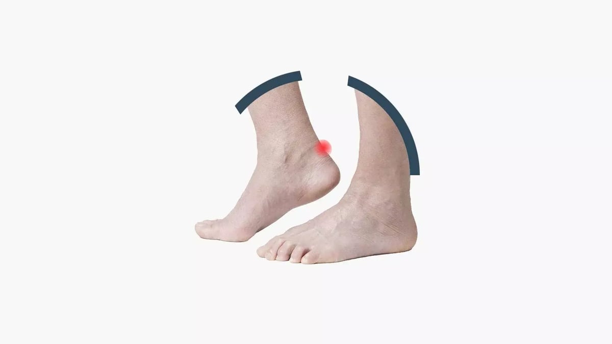
When the tendon becomes diseased at its insertion point in the Achilles region, the resulting inflammatory condition and painful symptoms are essentially caused by mechanical stress.
To simplify, we should imagine the tendon rubbing against the retroachilleal bursa (a tissue whose function is to reduce stress caused by shearing forces), which in turn rubs against the heel bone, which in turn rubs against the surrounding soft tissues, which themselves rub against the sock and the shoe.
We should picture a chain of shearing forces that produce friction and inflammation in the Achilles tendon.
This is why the athletes most affected by these issues are endurance sport athletes (middle-distance runners and marathoners), and those who use boots or footwear that increase posterior friction during their activity (such as skiers and football players).
In everyday life, the frequent and continuous use of heavy safety shoes can also contribute to the development of this condition.
Of course, there are various features that help define different subcategories of this pathology:
- Retroachilleal exostosis
- Intratendinous calcifications
- Retroachilleal heel spur
Insertional Achilles Tendinopathy: Diagnosis
Diagnosis is clinical. In this case, skeletal imaging completes the diagnostic process.
Often, a weight-bearing X-ray of the foot (particularly the lateral view) is sufficient.
In uncertain cases, it is preferable to use a second-generation imaging test, such as a Weight-Bearing CT scan, which provides a three-dimensional view of the heel bone and the Achilles tendon insertion under load. This offers a more comprehensive image while also correlating with functional weight-bearing conditions.
Insertional Achilles Tendinopathy: Conservative Treatments
This is the first strategy to pursue. Physical therapies play a key role.
The goal is to reduce local inflammation, as inflamed tissues typically have greater volume and are hypersensitive to pain.
The purpose of the therapies is to increase microcirculation, promote the removal of inflammatory byproducts, and by reducing inflammation, decrease the volume of tissues affected by the condition.
The most commonly used therapies are:
- Focused shockwave therapy
- Tecar therapy and laser therapy
Though their mechanisms of action differ, their objective is the same: to reduce inflammation and stimulate microcirculation.
Physical therapies should be accompanied by manual therapies and physical activity that helps improve tendon elasticity. The ideal exercises are eccentric loading exercises for the Achilles tendon (basically, stretching against resistance).
Personally, I never prescribe silicone heel lifts, which might have the benefit of slightly reducing mechanical stress on the tendon by inducing a mild equinus position, but undoubtedly have the negative effect of reducing the internal shoe volume and increasing the stress caused by shearing forces.
.
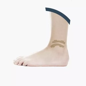
Non-Insertional Achilles Tendinopathy

The tendon can become diseased approximately two to three centimeters above its insertion on the calcaneus, leading to a characteristic hourglass-shaped deformity of the tendon.
Non-insertional Achilles tendinopathy, also known as Achilles tendonitis or tendinosis, is a condition that can have a subtle onset. The pain is not constant, at least in the early stages.
However, this condition carries a risk of Achilles tendon rupture. This risk has been linked to poor vascular supply to the tendon.
In fact, the condition typically occurs midway between the tendon’s insertion on the calcaneus and the muscle-tendon junction—that is, in the anatomical region farthest from the tendon’s vascular and nutritional support.
It most commonly affects patients aged 30 to 50, though it’s not uncommon even in people in their thirties. Early diagnosis is key to managing and minimizing the risk of a complete tendon rupture.
Non-Insertional Achilles Tendinopathy: Surgical Treatment
.webp?width=1200&height=675&name=f-usu-operatoria-desktop-min.jpg%20(1).webp)
When conservative treatments and regenerative medicine fail to provide the desired results, surgery becomes necessary to achieve a dual goal:
alleviate the patient’s symptoms, effectively resolving them, and
prevent a potential Achilles tendon rupture.
There are two main categories of surgical procedures for non-insertional Achilles tendinopathy:
- Surgery on the Achilles-plantar complex, often combined with regenerative medicine, depending on the goal:
- to stimulate the biological healing process of the diseased tendon
- to reduce biomechanical stress on the affected tendon
- Tendon transfer surgeries
Techniques to Stimulate the Biological Healing Process
Historically, the best known procedure is tendon scarification, which involves open cleaning of the tendon combined with small cuts to induce regeneration.
The real risk of this solution is that it damages the tendon more than the body is able to regenerate. For this reason, the natural evolution of this technique is Achilles stripping, a minimally invasive solution that, through four small 'small holes', aims to promote the detachment of the peritenonium (the sheath surrounding the Achilles tendon, which is richly innervated and vascularised) from the tendon, inducing deafferentation of the nerve fibres that transmit pain and inducing bleeding and, therefore, regeneration.
Based on the body's autologous regenerative capacity, stripping can be implemented with regenerative medicine techniques (an immunocentric revolution using mononuclear and mesenchymal cells taken from adipose tissue or bone marrow aspirate) to maximise the subject's regenerative potential.
Techniques for Reducing Biomechanical Stress on the Affected Tendon
There are several minimally invasive technical solutions: sectioning of the tricipital fascia according to Strayer, Bauman or Barouk.These techniques involve small incisions (less than 2 cm) in different locations, but with the common goal of releasing the Achilles-plantar complex.
These solutions can be combined with Achilles stripping and regenerative medicine.
TENDON TRANSFER SURGERY:
There are two different options:
- transposition of the flexor hallucis longus;
- use of the semitendinosus.
The semitendinosus tendon is taken from the ipsilateral knee, as is customary in anterior cruciate ligament surgery, without compromising knee function.
When using this technique, three small incisions are made, two proximal to the pathological area and one at the level of the calcaneus. With this type of tendon transposition, the semitendinosus is used to bridge the lesion in order to mechanically unload it. This controls the symptoms and reduces the risk of rupture.
It is an excellent, minimally invasive technique, with the limitation, however, of providing mechanical rather than biological support to the diseased tendon.
For this reason, transposition of the flexor tendon of the big toe is now much more widely used to supplement the Achilles tendon.
This is a surgical technique that can be performed endoscopically or through a small incision behind the tibial malleolus.
The flexor tendon of the big toe is isolated and transferred to the calcaneus. This technique provides biomechanical and biological (vascular) support for the tendon, controlling symptoms and reducing the risk of injury.
This procedure can be performed on an outpatient basis or with an overnight stay in hospital. Immobilisation with a brace is required for about 3 weeks, but weight-bearing is allowed immediately.
A return to normal daily activities with full weight-bearing is expected within 2 months, and a return to competitive sports approximately 4-6 months after surgery.
For further information, in this article we have discussed the main differences between insertional and non-insertional tendinopathy.
ACHILLES TENDON RUPTURE
With the exception of rare cuts, Achilles tendon rupture is always the final expression of a chronic degenerative process.
Patients often describe precisely the moment when the injury occurred.
However, in these cases, it is best to imagine the Achilles tendon as a frayed rope in which the final mechanism of injury plays a very minor role.
The rupture is an expression of the degenerative disease.
It generally affects patients between the ages of 40 and 60, but it is not uncommon in athletes in their thirties.
Patients typically describe a sharp pain, associated with the sensation of being “kicked from behind”.
In these cases, a completely conservative treatment that does not require surgery can be envisaged. However, this involves a series of casts (above the knee) and a period of immobilisation lasting approximately 90 days.
This is why, in patients with an active lifestyle, surgery is the fastest and most efficient way to recover after a complete Achilles tendon rupture.
The injury can be diagnosed clinically alone. Ultrasound is the first-line imaging test to confirm the clinical diagnosis.
The role of MRI (magnetic resonance imaging) is to establish the extent of retraction of the lesion stumps in order to identify the best technique.
In cases of minimal diastasis between the two lesion stumps, there are now minimally invasive surgical solutions that, even without incision or with a minimal incision (2-3 cm), allow realignment of the lesion stumps and excellent, rapid restoration “ad integrum”, minimising invasiveness.
For a more complete description of these treatment options, please refer to the specific article on Achilles tendon rupture.
In cases where the rupture has caused a diastasis of the stumps (more common in cases of chronic Achilles injuries, i.e. diagnosed late, more than 20 days after the event)
or in cases where the rupture occurs at the level of the myotendinous junction (i.e., a high injury), the surgical solution of choice for my team is tendon transposition of the flexor hallucis longus.
This avoids major injuries and uses a healthy tendon (the flexor hallucis longus that is transposed) as a tool for healing the injury.
This choice minimises the invasiveness of the operation and promotes a quick return to sporting activity.
However, it should be remembered that, regardless of the surgical choice, the recovery process requires immobilisation for approximately 40 days.
Our protocol differs from the traditional one, as it involves immediate immobilisation at 90° to promote early rehabilitation.
Full weight-bearing is allowed after approximately 2 months, and driving after a period ranging from 2 to 3 months.
Competitive sports are allowed after a period ranging from 6 to 9 months.
Obviously, in these cases, regenerative medicine plays a synergistic role in stimulating the regenerative processes initiated by surgery.
Muscle injuries: calf tear or strain
WHERE?
Muscle injuries can affect the deep layer (soleus muscle) or superficial layer (medial and lateral gastrocnemius muscles) and can be incomplete (strains) or complete (muscle tears). The patient reports pain in the calf area, which in the case of major tears may be a burning pain, comparable to an “amplified” cramp, and may be followed by the appearance of a bruise (haematoma).
Diagnosis
The diagnosis is clinical, but ultrasound and MRI scans are obviously important to complete the diagnosis, study the type of injury and monitor the healing process over time.
Cure
Treatment is conservative. In the early stages, therapeutic attention is focused on reducing the haematoma and preventing the injury from “worsening”. Physical therapies may be useful (laser therapy and Tecar therapy), but it is important to avoid hyperthermia, which can induce calcification at the site of repair.
The use of regenerative medicine is not recommended in most of these cases, at least in the acute phase.
During the rehabilitation phase, it is important to strengthen the muscles eccentrically in order to restore muscle strength and elasticity and protect the repair area.
Myotendinous injuries: Achilles tendon rupture
What are they?
Mytotendinous injuries are injuries in which the patient feels a sharp tear in the middle of the leg, comparable to the sensation of being kicked from behind.
The disability is significant and comparable to an Achilles tendon rupture, with a larger haematoma.
Diagnosis
The diagnosis is clinical. Thompson's test (squeezing the calf while the patient is lying prone and observing the presence of corresponding plantar flexion of the foot) and Vallum's sign (alteration of the normal anatomical profile with the presence of a local depression) are unequivocal. However, it can be difficult for the specialist to distinguish between a tendon rupture and a rupture at the myotendinous junction. This has an impact on the subsequent treatment.
That is why I believe that magnetic resonance imaging plays an important role in such cases: it allows the lesion to be located precisely!
Cure
In this case, the question to ask is: ‘Is it possible to heal conservatively, without surgery?’ The answer is yes.
Healing is a ‘restitutio ad integrum’; in other words, without surgery, will you return to how you were before? The answer is no.
Finally, conservative treatment requires long periods of immobilisation, often exceeding 90 days.
This is why treatment for this injury can be conservative, but in an active patient (not only in a patient with high functional demands or a top athlete), surgery is recommended.
This is especially true since tendon transpositions offer reliable, minimally invasive and fast treatment options.
In the past, these procedures were performed with large incisions and consequent significant surgical scars with the aim of repairing the injury, literally turning over a portion of the myotendinous fascia to regenerate the tendon and the broken myotendinous junction. These are moderately invasive procedures, with poor results in terms of functional recovery.
Today, this type of injury are treated with surgery that requires an incision of about 1.5 cm or endoscopy, taking a healthy tendon (flexor longus hallucis) from this incision, near the tibial malleolus, posterior to it, and using it as a new Achilles tendon and as functional and healing support for the injured Achilles tendon complex.
In short, instead of repairing degenerated tissue by simply putting a “patch” on it, the diseased functional unit is replaced with a healthy and well-vascularised tendon: the flexor longus hallucis.
In experienced hands, this procedure takes about 20 minutes and allows immediate weight-bearing, protected by a brace for 3 weeks, a return to driving and normal functions without braces after about 4-5 weeks, and a return to sports after about 3 months.
Prevention of Achilles tendon disorders
We are not talking about physical activity as a cliché or something we all know is important but which our daily routine has the ability to distract us from.
The Achilles tendon heals and ages with us.
The well-being of our body also coincides with the well-being of our tendons.
Our well-being is a cocktail of biochemical stimuli that our body modulates from both outside and inside. It is a cyclical and orderly rhythm, regulated by the sleep-wake cycle, which primarily governs our hormonal balance, along with other stimuli.
Physical activity is related to fatigue and our production of cortisol and other hormones.
This is why physical activity is important for our Achilles tendon and, first and foremost, the right physical activity is regular physical activity.
Setting achievable goals that are compatible with our daily routine and our level of well-being is essential, as is setting possible targets to achieve through improvement.
Therefore, the ideal physical activity for our tendons is one that we can plan regularly and that helps reduce our stress levels.
We are in line with modern bio-hacking theories, which many coaches and motivation and communication experts refer to.
Whatever activity you choose, it will be important to then focus on something specific for your Achilles tendon.
It is not true, for example, that running puts excessive strain on our tendons. However, if we are dedicated to running, it is useful to remember that our tendons need attention.
Regularly planning triceps (calf) stretches is essential to restore the correct elasticity of the tendon itself at the end of the chosen physical activity.
Stretching, however, also deserves consideration.
In fact, our muscles do not behave as isolated functional units.
Leibniz said: “a monad has no doors or windows”.
Well, our muscles are not monads, but are intimately related to the muscle groups with which they work (agonists and antagonists). Stretching and, if necessary, eccentric stimulation involving all the postural chains is therefore really effective.
In the case of the Achilles tendon, for example, it is important to also focus on the posterior muscles of the thigh (biceps femoris, but not only) and on the mobility of the pelvis. Finally, it is useful to also involve the antagonists, focusing on the tibialis anterior and extensors.
Nutrition, like physical exercise, contributes to well-being, but for years, the importance of various supplements or specific diets for the musculoskeletal system has been uncritically emphasised.
However, research has highlighted the presence of markers of well-being that predispose individuals to healing or reduce the incidence of systemic or local inflammatory diseases.
Among these, vitamin D and the presence of polyunsaturated fats are indicators.
Our centre specialising in Achilles tendons and the value of our research

My team, supported by the Scientific Directorate of IRCCS Galeazzi, where I worked for 10 years before becoming Head of Ankle and Foot Orthopaedics at Humanitas San Pio X, designed and conducted the first study described in Orthopaedics on the use of cells taken from adipose tissue as a regenerative stimulus for non-insertional Achilles tendon pathology.
In this study, we compared PRP with the stromal fraction of adipose tissue for the first time.
Our study was the first to describe this treatment as safe and effective.
This study formed the basis for further scientific research and the identification of better imaging methods for the Achilles tendon. To date, magnetic resonance imaging, with dedicated mapping, is an ideal tool for monitoring the evolution of non-insertional Achilles tendinopathy over time.
The results of these studies have been published in some of the most prestigious journals in the field (KSSTA, British Bulletin and Foot and Ankle Clinics).
This is an area of research to which my team remains passionately committed, convinced that regeneration is the key to reducing the invasiveness of surgical procedures and achieving ever greater patient satisfaction.