Skip to content

Ankle Osteoarthritis


Healing means acting, it is not a passive event.
– Caroline Myss –


Ankle Arthritis

Ankle arthritis is a chronic joint disease (arthropathy) that can affect any joint in the body—from the spine to the upper and lower limbs.

It is a degenerative condition caused by the gradual breakdown of the cartilage that normally covers and protects the joint surfaces. As the cartilage wears away, the underlying subchondral bone becomes exposed, leading to progressive changes in the shape and function of the joint.

Therefore, it is not a disease of cartilage alone, but of the entire joint:

  • skeletal structures;
  • ligaments;
  • capsular structures,

and even the muscles, connected with joint movement, are involved.

When it comes to the ankle specifically, Dr. Steven Raikin—orthopedic surgeon at the Rothman Institute in Philadelphia and member of the AOFAS—has recently demonstrated that ankle arthritis has a profoundly negative impact on quality of life, comparable to that of hip osteoarthritis and, in some cases, even more debilitating than knee osteoarthritis.
Unlike other forms of arthritis, which typically correlate strongly with aging, ankle arthritis does not follow this trend. Its prevalence is not primarily age-related, making it a unique and particularly challenging condition to manage.

Unlike the knee or hip, the ankle joint is what we doctors call "highly congruent"—meaning the surfaces of the joint fit together with exceptional precision.
On the one hand, this anatomical congruence acts as a protective factor against degenerative arthritis. On the other, it explains why even a single traumatic event that disrupts the joint’s anatomy can be enough to trigger significant arthritic changes.
This is why approximately 70% of ankle arthritis cases have a post-traumatic origin.
Patients who have sustained fractures involving the malleolus, tibia, fibula, talus, or calcaneus—even when these fractures have healed, whether perfectly or not—are at risk of developing ankle arthritis.
To this group, we must also add those with chronic ankle instability due to ligament injuries or structural deformities.
In the remaining cases, ankle arthritis is linked to chronic systemic inflammatory conditions, such as rheumatoid arthritis, or other diseases that can lead to joint damage, such as hemophilia.

What is osteoarthritis of the foot?

It is a pathological condition that occurs when osteoarthritis is not limited to the ankle but also involves other joints of the foot, such as those of the tarsus. This situation can result from trauma, congenital malformations or functional overload and typically manifests with debilitating symptoms.
The main signs of osteoarthritis of the foot include:

  • progressive joint deformity;
  • chronic and persistent pain, which may intensify during movement;
  • difficulty walking and performing daily activities.

To improve this condition, it is essential to take prompt action. In the early stages, conservative therapies such as physiotherapy, the use of orthopaedic insoles or anti-inflammatory drugs can reduce pain and improve function.
In more advanced cases, personalised surgical procedures may be used to restore the biomechanical balance of the foot.

The causes of ankle osteoarthritis

artrosi caviglia diagnosi


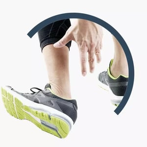
Ankle osteoarthritis differs from other forms of osteoarthritis in its particular clinical profile: the typical patient is young, aged between 30 and 50, with a history of trauma, such as fractures (e.g., ankle, tibia and fibula, tibial pilon, malleolar, bimalleolar or trimalleolar fractures).
Let me explain. In general, the onset of osteoarthritis is directly related to age. Unlike other joints, the ankle has extremely congruent joint surfaces that fit together almost perfectly, like a jigsaw puzzle. This feature reduces the likelihood of age-related wear and tear and explains why ankle osteoarthritis is often a consequence of previous trauma.

Approximately 70% of cases of ankle osteoarthritis are post-traumatic in origin: they result from a fracture or dislocation of the ankle itself, which can affect the entire lower limb.

This explains why ankle osteoarthritis usually affects young people, who are more prone to high-impact trauma, due to its post-traumatic nature.
Even when treated correctly, these traumas can cause a loss of joint congruity, which in turn is responsible for the development of osteoarthritis in the joint.
The post-traumatic nature of ankle osteoarthritis has another peculiarity: malalignment, often associated with complex, displaced or exposed fractures, which are difficult to reduce and typical of high-energy trauma. Despite rigorous treatment, these fractures can cause joint deformities, such as varus.
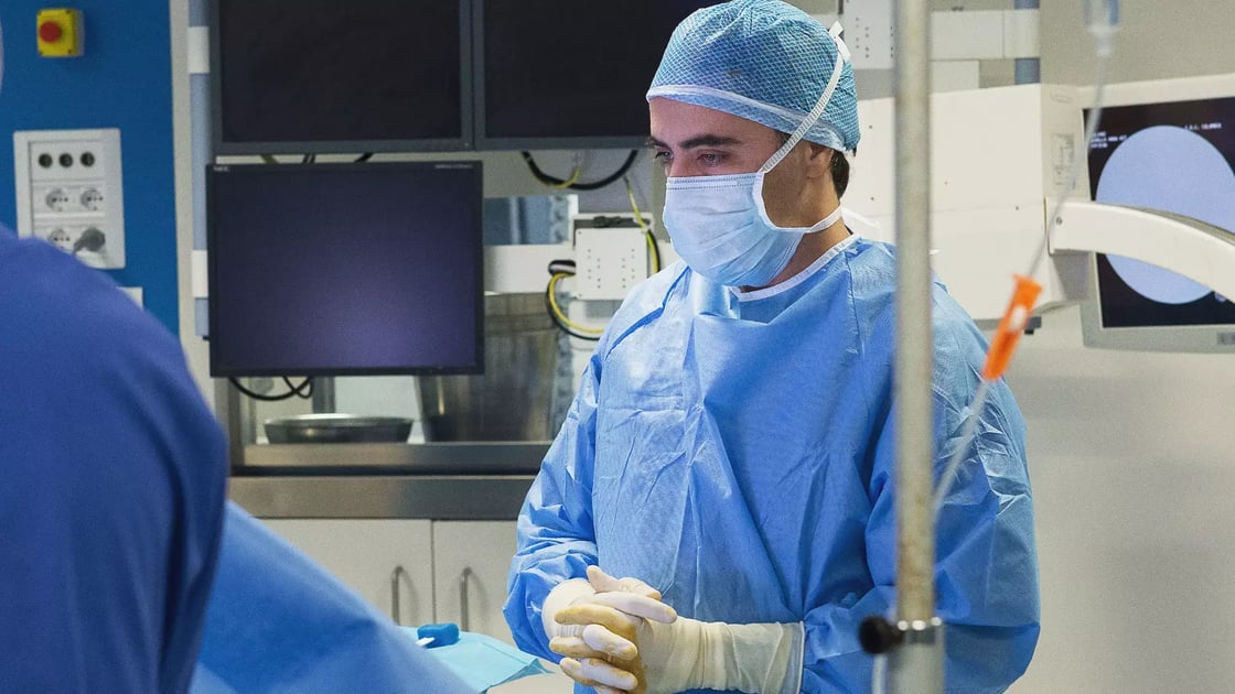
Unlike hip and knee prosthetics, which in many cases involve surgery on well-aligned joints or joints with slight misalignments, ankle surgery frequently involves significant deformities, which make surgery particularly complex.
For this reason, it is essential to rely on an experienced surgeon and specialised centres for the treatment of this condition.

Ankle Arthritis Symptoms


The main symptom of ankle osteoarthritis is pain, often accompanied by joint stiffness.
 

Pain

Pain is the first sign that something is not working as it should. This is a consequence of the deterioration of the joint cartilage and the misalignment often associated with it, which prevents the joint from working properly.
The deterioration of the joint cartilage and the misalignment often associated with it, common consequences of the osteoarthritic process, create a vicious circle of inflammation and progressive damage to the cartilage. 
This leads to pain during walking and certain joint movements.
At first, the pain is more acute after long periods of immobility, for example upon waking in the morning. As movement resumes during the day, these symptoms subside, although they may flare up at times.
In later stages of the disease, however, pain also occurs at rest, is deep, poorly localised, and is exacerbated by previous joint overload or changes in weather conditions.


Stiffness

Joint stiffness is another important feature of osteoarthritis, caused both by the formation of osteophytes, which are the body's attempt to stabilise the joint, and by muscle contraction, which gradually becomes less toned due to reduced movement. This slow and progressive loss of mobility does not improve without specific treatment.
The joint may appear painful on palpation, during passive movements and, of course, when walking. During movement, crepitus or cracking may also be heard due to the wear and tear of the joint cartilage, joint misalignment and/or the presence of osteophytes.

Puffiness

Slowly and progressively, ankle osteoarthritis leads to puffiness in the affected area, which may also be painful to the touch. The swelling is caused by inflammation and the build-up of synovial fluid, the fluid contained in the protective membrane of the joint that serves to “lubricate” the joint itself.

Deformation

Finally, it should be noted that due to its predominantly post-traumatic nature – 70% of cases, as mentioned above – the ankle may appear deformed and misaligned (not in axis). This is because reduction and synthesis procedures performed after an ankle fracture do not always restore the correct anatomy of the joint.
Deformity is another distinctive symptom of ankle osteoarthritis. A consequence of severe, displaced and sometimes exposed fractures related to high-energy trauma (e.g. road accidents, motorcyclists, sports), this deformity, typically in varus, complicates the overall picture, causing overload of certain joints and making them work incorrectly.
The deformity is therefore not only damaging to the ankle itself, but can also cause early osteoarthritis in neighbouring joints such as the subtalar joint and even the knee.

Diagnosis

Once the disease has been diagnosed and the symptoms carefully evaluated, the next step is to proceed with the appropriate diagnostic tests.

A simple X-ray is usually sufficient to reveal the typical signs of osteoarthritis, including:

  • Narrowing of the joint space
  • Remodeling of the subchondral bone
  • Formation of osteophytes (small bone spurs)
  • Presence of geodes (bone cysts)

If surgical treatment becomes necessary, more detailed assessments are recommended.

Since the ankle functions as part of the foot's biomechanics, its ability to function properly depends on the surrounding structures. For this reason, X-rays of both the foot and ankle are essential to evaluate alignment and determine what surgical interventions may be required to ensure a long-term stable implant.

I would also like to emphasize an important point: Contrary to popular belief, MRI does not provide more useful information than X-rays in this context. This is because MRI is performed with the patient lying down, without weight-bearing, which is a crucial factor in assessing ankle function.

In addition to standard imaging, I strongly recommend an X-ray projection developed by my colleague Donald I. Saltzman, performed with the foot inclined at a 20-degree angle. This simple technique allows for a more accurate analysis of the relationship between the calcaneus, talus, and tibia, offering a clearer understanding of the biomechanical alignment.
Finally, in planning for surgery, a CT scan of the ankle and hindfoot is also essential. It provides valuable information about bone quality, which plays a critical role in choosing the most appropriate surgical approach.


How do you Cure Ankle Arthritis?

dr. Federico Usuelli

Ankle osteoarthritis is an irreversible degenerative condition. While it can be managed to control symptoms, full recovery of a damaged joint is currently not achievable.

NSAIDs (Non-Steroidal Anti-Inflammatory Drugs) are effective for pain relief, but long-term use is discouraged due to potential side effects such as gastritis and ulcers. The use of opioids, like morphine, is also not recommended because of the risk of addiction. Similarly, cortisone injections carry risks including osteoporosis and infections.

Ankle osteoarthritis: conservative treatments

tecar terapia

In summary, pharmacological treatments can provide symptomatic relief during certain phases but do not offer a long-term solution.

Physical therapies

There are also physical therapies, such as InterX and Tecar Therapy, which can be even more effective when used together. These therapies improve the biology and function of the tendons and muscles involved in joint movement. In the early stages of arthritis, they can serve as a valid, repeatable option that may delay the need for surgery for a significant period.
Important: These therapies require the supervision of an experienced, qualified professional who collaborates closely with an orthopedic specialist focused on foot and ankle care.

Medicina rigenerativa: PRP

Based on my experience, I generally advise against intra-articular injections for the ankle. This is especially true for cortisone. Recently, new treatments such as viscosupplementation with hyaluronic acid and its derivatives, or injections of platelet-rich plasma (PRP), have been proposed—but without strong scientific evidence supporting their use. The results so far are mixed and often inconsistent.

How can ankle osteoarthritis be prevented?

Ankle osteoarthritis is symptomatic in most cases, but not all cases ‘necessarily’ require a prosthesis.
Prevention means:

  • optimal trauma management
  • treatment of deformities and prevention of their progression (joint preserving surgery)
  • cartilage treatment
  • Regenerative Medicine


Preventing the progression of osteoarthritis and preserving ankle function is possible with targeted and timely interventions.


Ankle Surgery and Replacement

In the three images above, from left to right, you can see: a healthy ankle, a fractured ankle, and a prosthetic ankle.

There are two main surgical options for treating ankle arthritis: arthrodesis (joint fusion) and arthroplasty (joint replacement).

1.Artrodesi

Arthrodesis is the surgical fusion of a joint. Here, we focus on ankle fusion.
This procedure can be done either through an open incision or arthroscopically. In both cases, the goal is to achieve complete fusion of the ankle joint at a 90-degree angle.
However, this means sacrificing all ankle movement. Additionally, it places increased stress on the surrounding joints, which then bear more load and face a higher risk of developing osteoarthritis.
For younger patients, ankle arthrodesis carries the risk of osteoarthritis spreading to other foot joints. Over time, this may require further fusion surgeries, resulting in a progressively stiffer and less flexible foot.

2.Protesi di caviglia

Ankle replacement surgery is currently the gold standard treatment for ankle osteoarthritis. It involves the total replacement of the degenerated joint with an implant that is able to reproduce movement and is as close as possible to its physiology.
Compared to patients with hip or knee osteoarthritis, those affected by ankle osteoarthritis can be much younger and, as such, have greater mobility needs in their daily lives. This factor is no longer a contraindication for prostheses, thanks to new-generation implants.
Ankle prostheses are a reliable solution thanks to the increasingly advanced materials used and prosthetic designs aimed at reproducing the anatomy and biomechanics of the ankle as accurately and physiologically as possible.
Even in cases of pre-existing deformities, prostheses are now recommended. In such cases, it is essential that the surgeon has extensive experience in ankle prosthetics and reconstruction techniques.
If you would like to learn more about this topic, please read: ‘Ankle prostheses: 12 important facts for patients’.

Contraindications and new techniques

Contraindications and new techniques
The only contraindication to prosthesis, to the extent that ankle arthrodesis is recommended, is the lack of bone stock (ascertainable by CT scan), i.e. the lack of bone reserve on which to support the implant.

In patients with insufficient bone stock or those suffering from peripheral neurological disorders (drop foot), arthrodesis, i.e. fusion of the ankle, which is locked at 90° to the leg, remains the only option.

When searching for information on the internet about prosthetic techniques, you may come across a variety of opinions against this type of surgery. This erroneous school of thought has taken root due to the old prostheses used more than 15 years ago.

Today, cases in which ankle prostheses cannot be performed are increasingly limited thanks to new prosthetic designs, which allow for significant bone savings, giving the surgeon the possibility to position a prosthesis even in situations where the bone stock is very reduced.

Looking to the future, there is a growing trend towards a technique known as “resurfacing”, which involves reducing the volume of prosthetic implants and developing prosthetic designs that are increasingly similar to the original anatomy of the ankle.

This is possible thanks to materials that can reproduce the physical characteristics of bone, including its porosity, rigidity and elasticity.

For example, the introduction of a material such as Trabecular Metal (tantalum subjected to a special process) now allows a resurfacing procedure with smaller, more anatomical implants, which significantly speeds up recovery time. This implant is inserted through a lateral incision, rather than an anterior incision as is usually the case.

In Italy, I am currently the surgeon who has performed the highest number of operations and follow-ups* using the resurfacing technique. It is a revolutionary and reliable choice that offers great advantages but is currently reserved for selected clinical cases.

With over a hundred operations performed using the “Hintegra” (Mobile-Bearing) prosthesis and over three hundred cases with the TM-Ankle Zimmer-Biomet prosthesis (Fix-Bearing or Resurfacing with lateral access), I am a unique point of reference in Italy for the treatment of ankle osteoarthritis. In addition, my experience is supported by an extensive body of scientific literature published on PubMed.

Useful article for further reading: “Ankle prostheses: the results of our surgical technique in JBJS America”.

Recovery

Before surgery, the patient undergoes pre-admission, which includes blood tests, a final orthopedic evaluation, and an anesthetic assessment.
The anesthesia used is usually a combined technique that provides prolonged numbness in the leg—often lasting until the next morning—to minimize the need for pain medications.
Hospitalization typically begins on the day of surgery or the day before, and usually lasts between 2 to 4 days. The patient is discharged once the surgical wound is healing well and pain is manageable without daily medical supervision.

Returning on Field

post artrosi caviglia riabilitazione

Ankle replacement surgery is becoming increasingly common, but it requires careful follow-up and a delicate post-operative course focused on ensuring proper osseointegration before early mobilization.
My patients are immobilized with a cast or brace for six weeks, although weight-bearing on the foot is generally allowed after three weeks.
It is crucial that patients attend weekly check-ups to monitor skin healing, typically occurring between 3 and 5 weeks after surgery.
Between 3 and 6 weeks, the first standing X-rays are performed.
Once the cast or brace is removed, walking rehabilitation becomes essential. I usually recommend hydrokinesitherapy (water-based walking exercises), stretching of the triceps surae muscle, and later proprioceptive training.
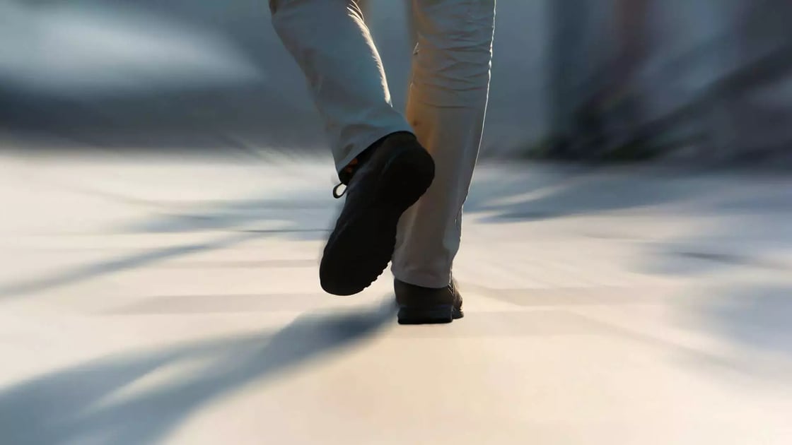
Most patients regain self-sufficiency about two months after surgery, can resume driving after 3 to 4 months, and achieve full post-operative satisfaction around 6 to 8 months.
These recovery times can be significantly shortened with new resurfacing techniques, although each case must be evaluated individually.

Scientific Research and Ankle Arthritis

My dedication to preventing and improving surgical treatments for ankle arthritis is reflected in my daily commitment to study and research. To explore these topics in greater depth and to appreciate the innovations I have contributed to on an international level, I invite you to visit the “Scientific Papers” section.

More